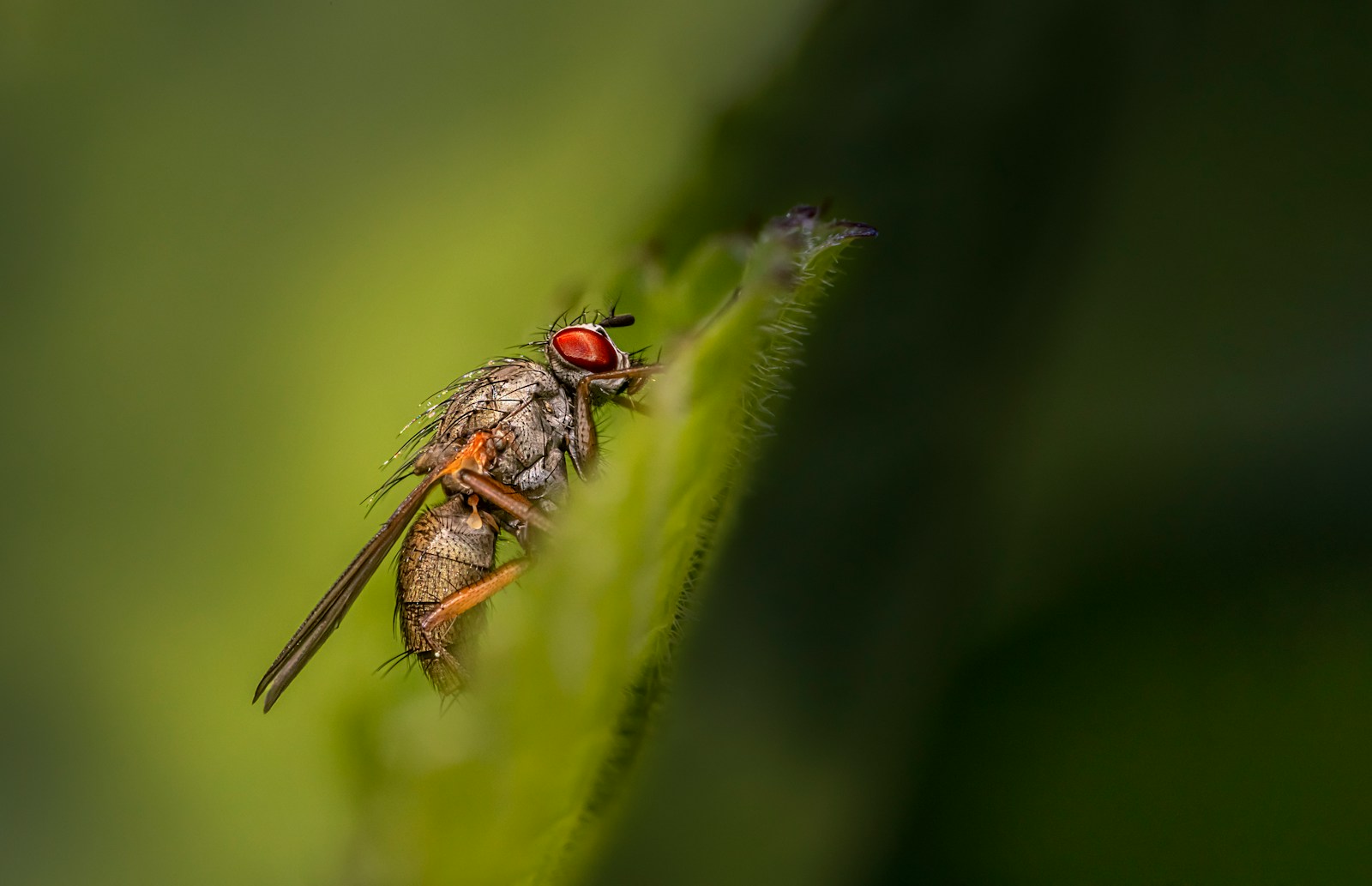
The human brain consists of billions of neurons. These neurons are connected by synapses, where information is passed from one neuron to another. Understanding how neurons connect and communicate can reveal how the brain controls behaviour. However, reconstructing these connections for an entire brain has been challenging due to technological limitations. Recently, a team of scientists created a detailed brain map of conection using electron microscopy techniques using the brain of Drosophila melanogaster (the common female fruit fly). Although fruit flies have smaller brains than humans, they still engage in complex behaviours, such as flying, social interaction, and memory formation. Mapping the fruit fly brain can help scientists understand how neurons work together to process information and generate behaviour.
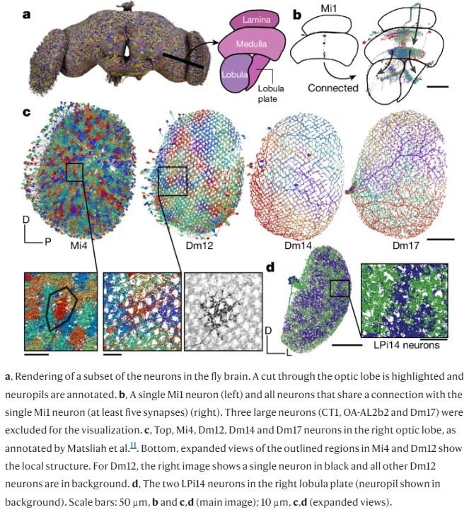
Researchers used electron microscopy to image the entire brain of an adult female fruit fly. Electron microscopy provides extremely detailed images, which allowed the researchers to see individual neurons and their connections (synapses). They segmented these neurons and synapses using computational techniques and built a map that shows how each neuron connects to others across different brain regions.
The connectome reveals several important features of how the brain processes information.
Neuron classes in brain map
The researchers found that the brain contains three main types of neurons:
Intrinsic neurons: These are confined entirely within the brain. They make up the majority of neurons and are involved in processing information within the brain itself.
Afferent neurons: These neurons bring sensory information into the brain from the outside world. They are mostly located outside the brain and connect through nerves.
Efferent neurons: These neurons send signals from the brain to other parts of the body, like muscles, glands, and other organs.
Synapse Distribution: The study identified that synapses (connections between neurons) are not evenly distributed. Some neurons have thousands of synapses, while others have fewer. The density of synapses in the fruit fly brain is much higher than in the mammalian cortex, with 7.4 synapses per cubic micrometer.
Brain connectivity
The researchers developed a “projectome,” which maps the connections between major brain regions, called neuropils. The projectome shows how sensory information flows from input neurons (like those involved in vision and smell) to output neurons (like motor neurons that control movement). This map provides insights into how the brain processes information on a larger scale.

Motor pathways: A detailed analysis of the connections between sensory neurons (photoreceptors) and motor neurons showed how the brain integrates sensory input to produce movement. The study highlighted pathways involved in sensorimotor behaviours, such as flying and walking.
Asymmetry and cross-hemispheric connections: The brain has two hemispheres, and most connections occur within one hemisphere. However, the researchers also discovered neurons that connect the two sides of the brain. These connections allow for coordination between the two hemispheres, which is essential for behaviours that require symmetry, like movement and balance.
Visual and sensory neurons: The optic lobes, responsible for visual processing, contain a large number of neurons. These neurons project into other brain regions, influencing decision-making and movement based on visual input. The study identified 8,053 visual projection neurons (VPNs) that send information from the eyes to the central brain.
Comparisons with other species: The fruit fly connectome provides a model for comparing brain connectivity across different species. Researchers can now compare this connectome with the simpler connectome of Caenorhabditis elegans (a worm) and the developing connectome of vertebrates like mice. This will help scientists understand how neural circuits evolve to handle more complex behaviours.
Function
The connectome allowed the researchers to analyze how specific neurons contribute to different functions. By tracing the pathways from sensory neurons to motor neurons, the researchers could make predictions about the role of certain neurons in behaviour.
Ocellar pathway: The ocelli are small visual organs that help the fruit fly detect changes in light, especially useful for flight control and orientation. The study traced the pathway from the ocelli to neurons in the central brain and then to descending neurons that control motor actions. This pathway suggests that ocelli are involved in stabilizing flight and adjusting the fly’s orientation based on light levels.
Information Flow: By using a computational model, the researchers analyzed how information flows through the brain. They seeded the model with afferent (input) neurons and traced how information moves through different layers of the brain. The results showed that different sensory modalities (vision, taste, smell) have specific pathways that they follow, but all information converges in higher-level brain regions that make decisions or produce movement.
Neuron superclasses and network analysis: The connectome allowed the team to categorize neurons into superclasses based on their location and function. These superclasses included neurons that primarily receive input from the environment, neurons that process information within the brain, and neurons that send output to the body. The study found that the brain’s internal connections are more numerous than the external connections, meaning that the brain communicates more within itself than with the outside world.
Neurotransmitter predictions: The researchers predicted the neurotransmitters used by each neuron. This helped them understand whether certain neurons are excitatory (promoting action) or inhibitory (reducing action). For example, GABAergic (inhibitory) neurons tended to have more connections than other types of neurons. This balance between excitatory and inhibitory neurons is essential for controlling the brain’s activity and preventing overstimulation.
Implications for future research
The fruit fly connectome provides a comprehensive map of how neurons are wired in the brain, making it a valuable tool for future research on brain function and behaviour. Researchers can now use this connectome to test hypotheses about how the brain controls behaviors such as memory, decision-making, and movement by studying specific neural circuits.
The connectome also allows scientists to explore variability between individuals or species, providing insights into how genetic and environmental factors influence brain development and function. By comparing different connectomes, researchers can uncover patterns and differences in brain wiring that contribute to diverse behaviours.
Additionally, the connectome serves as a blueprint for modeling complex behaviors. By simulating brain activity in computational models, scientists can predict how neurons interact and respond to various stimuli, offering deeper understanding of brain functions.
Finally, the fly connectome sets the stage for cross-species comparisons. Researchers can now examine similarities and differences in brain wiring between flies, worms, and mammals, helping to uncover the evolution of neural circuits and providing a basis for understanding how complex behaviours arise across different species.
Conclusion
The complete connectome of an adult female Drosophila melanogaster brain represents a significant step forward in neuroscience. It provides a detailed map of how neurons are connected and offers insights into how the brain processes information and controls behaviour. This resource will guide future research in understanding brain function, behaviour, and neural computation, not just in fruit flies but also in other species. The open nature of the data means that researchers from around the world can access and contribute to the ongoing exploration of the fly brain.
References
Dorkenwald, S., Matsliah, A., Sterling, A. R., Schlegel, P., Yu, S., McKellar, C. E., Lin, A., Costa, M., Eichler, K., Yin, Y., Silversmith, W., Schneider-Mizell, C., Jordan, C. S., Brittain, D., Halageri, A., Kuehner, K., Ogedengbe, O., Morey, R., Gager, J., . . . Murthy, M. (2024). Neuronal wiring diagram of an adult brain. Nature, 634(8032), 124–138. https://doi.org/10.1038/s41586-024-07558-y


